Ocular Coherence Tomography (OCT)
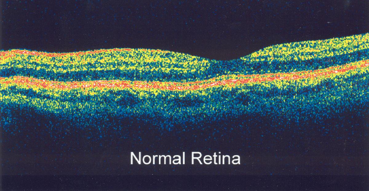
The latest advance in eye imaging is known as Ocular Coherence Tomography (OCT). OCT is a non-invasive imaging technology of the retina, similar to ultrasound testing except it measures light waves rather than sound.
OCT is useful in diagnosing or evaluating many eye conditions, including:
– macular hole
– macular pucker
– macular edema
– age-related macular degeneration
– glaucoma
– diseases of the retina
– diabetic retinopathy
– vitreous traction
– optic nerve fiber changes
OCT has evolved over the years to allow incredibly high resolution cross sectional images of the retina, iris, and cornea. The application is primarily directed toward the diagnosis and treatment of glaucoma and macular degeneration that has led to the newest version of the technology called OCT Angiography. OCT-A, which we are proud to say we now have available, can scan the blood vessels down to the smallest micro – capillaries in the entire retina to determine either the lack of blood flow to retina tissues, or destructive blood vessel formation as might be seen in wet macular degeneration or severe diabetic retinopathy. This imaging allows visualization months to years prior to conventional diagnostic methods that primarily rely on the presence of symptoms. This is obviously too late.
A full complement of diagnostic equipment is available including cornea topography to determine the shape in health of a cornea, 2 different visual field testing instrument to map any visual loss in the peripheral vision and several digital camera systems that image pathology in the anterior segment of the eye including the eyelids and lashes, cornea lesions, tear film stability and anomalies of the conjunctiva, sclera and iris.